Flow cytometry is a popular cell biology laboratory method. It uses a laser for rapid analysis, quantification, and sorting of a suspension of live cells. In a matter of nanoseconds, profiles of the cell population’s properties are acquired, including size and granulation. This is based on the characteristic pattern of light refracted from the cells. It is then possible to purify the cell population by sorting them into distinct channels depending on their distinct properties.
 Carl Dupont | Shutterstock
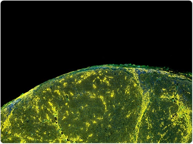
When a more in-depth investigation of a cell population is required, antibodies tagged with fluorescent dyes are used to identify the presence or absence of particular cellular markers. Detecting specific proteins (such as CD markers) in a cell population may help to determine cell types, and this is known as immunophenotyping.
What are fluorochromes?
Fluorescent markers are known as fluorochromes, a term that is used interchangeably with fluorophore. Upon absorption of light from a laser, they become ‘excited’ and emit light at a longer wavelength to return to their original ‘ground’ energy state, which is known as fluorescence.
Each fluorochrome absorbs and emits light at a specific range of wavelengths. The emitted, longer wavelength of light is ideally a different color to the laser for easy detection of fluorescence. For example, the most widely used fluorochrome, Fluorescein isothiocyanate (FITC), has a peak absorbance of 494nm and a peak emission of 512nm corresponding to the colors blue and green.
Exciting fluorochromes at their peak absorbance (rather than at the bottom end of their absorbance spectrum), enables more intense light emission. Although fluorochromes have specific wavelength spectra, they are often overlapping. Therefore, when using multiple fluorochromes, it is important to choose those that are non-overlapping. This allows differentiation of the cell features they are highlighting.
Stokes shift
Another important point to consider when choosing a fluorochrome is the Stokes shift: the difference between the wavelength of the exciting photon (the laser) and the emission photon (from the fluorescing cell). A large Stokes shift is desirable to distinguish between the different colors of the exciting and emitting photons.
Tandem dyes
Tandem dyes are made by covalently binding two different fluorochromes. One becomes the donor and the other an acceptor. The donor is ‘excited’ and emits light that is absorbed by the acceptor, due to Fluorescence Resonance Energy Transfer (FRET). For this to occur, the two fluorochromes require overlapping emission/absorption spectra.
One advantage of this method is that it further increases the Stokes Shift for more distinguishable colors. It enables the use of several fluorochromes in a panel, producing many colors for analyzing multiple parameters. One limitation of tandem dyes is that they are highly sensitive and degrade easily, especially when exposed to light, resulting in reduced fluorescence. It is also necessary to monitor the pH environment of all fluorochromes, as differing pH levels change their brightness.
Quantum dots
Small nanocrystals (2-20 nm), known as quantum dots, are an alternative to fluorochromes. The wavelength emitted depends on the size of the quantum dot. They have much narrower emission spectra compared to standard fluorochromes, reducing the chance of overlapping colors when multiple must be used.
Further Reading