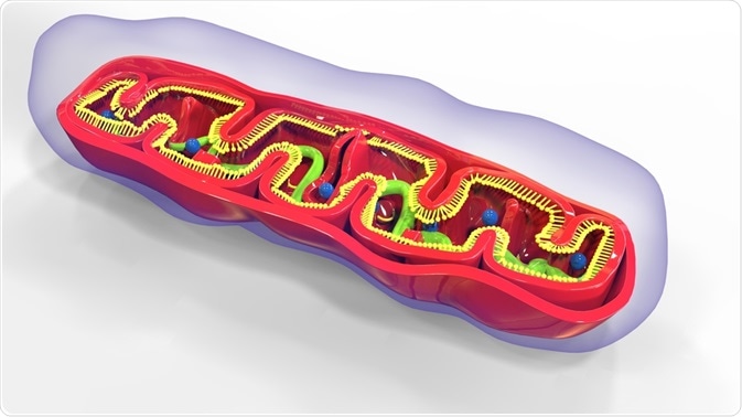
There are a total of 5 multi-subunit complexes in the inner membrane of the mitochondria, all of which are involved in the process of oxidative phosphorylation that precedes ATP production. Of these, 13 subunits are encoded by mitochondrial genes (mtDNA). The mitochondrial genome is therefore vital for the production of cellular energy. Defects in this mechanism may result in a litany of mitochondrial diseases.
Skip to:
 MicroScience | Shutterstock
The mammalian mitochondrial genome comprises 2 rRNAs, 22 tRNAs, and numerous mRNAs, which encode the 13 polypeptides required for oxidative phosphorylation. The entirety of the mitochondrial genome is transcribed to produce just two transcripts: heavy (H) and light (L), so-called based on their positions in a cesium chloride density gradient.
Extensive processing of the transcripts is required to produce fully functional RNA components. These include cleaving of the primary transcript, further polymerization and chemical modifications such as adenylation and aminoacylation.
The Heavy and Light Regions of mtDNA
The heavy and light regions contain two classes of promoters: the heavy strand promoters (HSPs) and the light strand promoters (LSPs), respectively, which provide the origin of translation. The LSP controls the transcription of eight tRNAs and a mitochondrial gene MT-ND6.
There are two promoters proposed to be associated with the heavy strand: the first, HSP1, governs the expression of tRNAPhe, tRNAVal, 12SrRNA and 6S rRNA. The second promoter, HSP2, produces a transcript that encompasses almost the whole of the mitochondrial genome.
The existence of two promoters was popularized by the work of Ojala et al., who noted that the 12S mitochondrial rRNA transcript (MT-RNR1) was 100-fold more populous than most mRNAs despite being encoded on the same strand. Two explanations were postulated: (1) the presence of two HSPs, or (2) early termination of the 16S rRNA (MT-RNR2).
The two-promoter theory is therefore thought to account for the high abundance of the tRNA transcripts. Their existence has been confirmed, and the activity of both HSP1 and HSP2 can be observed in vitro.
Interestingly the in vivo and in vitro transcription start sites of HSP2 are not congruent and this has favored the theory of a single promoter. The evidence comes from experiments employing mitochondrial termination factor 1 (MTERF1) knockouts.
MTERF1 exclusively stimulates HSP1 and not HSP2 activity. When it is knocked down, however, cells do not display changes in the relative abundance of HSP1 and HSP2 transcripts as would be expected. To conclude, there is insufficient evidence available to suggest that two promoters exist on the heavy strand.
Mitochondrial transcription is a three-step process as with genomic DNA transcription: initiation, elongation, and termination.
Transcription Initiation
The polymerase POLRMT drives human mitochondrial transcription. It is structurally similar to both the T3 and T7 bacteriophage RNA polymerases, and all three have a conserved catalytic core at the C-terminus. POLRMT differs in that it also possesses duplicated domains, the pentatricopeptide repeat (PPR), which dictates site-specific interactions.
Compared to the T3 and T7 bacteriophage polymerases, POLRMT requires auxiliary proteins to recognize the promoter. These are the DNA-binding protein mitochondrial transcription factor A (TFAM) and mitochondrial transcription factor B2 (TFB2M).
TFB2M is a gene duplication product which controls local ‘melting’ of the DNA necessary for transcription. The other product of duplication is a methyltransferase TFBM1, the catalytic domain of the methyltransferase is also maintained in TFBM2 although its predominant function is DNA ‘melting’. The transcription initiation complex forms at both promoters on the heavy and light strands.
The process begins when TFAM binds to its high-affinity sites upstream of HSP1 and LSP. This causes a sharp 180° bend in the DNA molecule. This 180° bend at the LSP is thought to be essential for the correct positioning of the mitochondrial transcription machinery with regards to the transcriptional start site (TSS).
The TFAM-mtDNA complex then recruits POLRMT, which is bound by its N-terminal extension to the C-terminal domain (CTD) of TFAM. This establishes the pre-initiation complex (pre-IC). The recruitment of TFBM2 induces DNA unwinding at the promoter and enables initiating nucleotides to be incorporated into the mtDNA/TFAM/POLRMT/TFBM2 ternary complex, thus beginning RNA synthesis. Following initiation, TFBM2 is dissociated to enable elongation to commence.
Transcription Elongation
The transition from initiation to elongation requires the transcription elongation factor (TEFM). In vitro, recombinant TEFM is observed to increase POLRMT processivity: the ability for an enzyme to catalyze reactions in a continuous manner without disengaging with its substrate.
This increased POLRMT processivity induced by TEFM stops G-quadraplexes from forming. These are nucleotide-based helical secondary structures that are rich in guanine. They may be formed from one, two or four strands and are further stabilized by cations, particularly potassium.
The formation of G-quadraplexes is predominant in the light strand LSP and inhibits continued elongation from the LSP at a specific region called the conserved sequence block 2 (CSB2). The resultant short RNA transcripts are implicated in priming DNA replication, a view corroborated by the presence of multiple RNA to DNA transition sites about CSB2.
The prevention of G-quadruplex formation by TEFM subsequently prevents premature termination at CSB2 and thus functions to switch the process of replication to transcription of the LSP-derived primary transcript. Structurally, the TEFM forms a sliding clamp around the mtDNA via its pseudonuclease core. Additionally, TEFM functions downstream of POLMRT and associates with POLMRT via its C-terminal domain.
Transcription Termination
The mechanism of HSP termination is unclear. It was previously hypothesized that termination is mediated by MTERF1. Recombinant MTERF1 mediates termination on both the heavy and light strands (bidirectional termination). It binds on the major groove of the mtDNA helix and induces a 25° bend, which partially unwinds it. This induces base flipping, or eversion, which stabilizes the binding of MTERF1 to the mtDNA, which facilitates its obstruction of the transcription elongation machinery.
More recent evidence contradicts this mechanism. The increased rRNA quantity was thought to be caused by this, however, knockdown of MTERF1 does not affect the levels of rRNA, suggesting their abundance is a consequence of their stability.
Termination of the LSP is not due to DNA bending induced by MTERF1. Instead, MTERF binds at the 3’-ends of the coding region of the mtDNA and prevents the encroaching replication fork from progressing, whilst simultaneously obstructing antisense transcription of the rRNA genes.
Primary Transcript Maturation
The resultant transcript is polycistronic, encoding several proteins. It is comprised of protein-coding sequences that are interspersed with mt-tRNAs. mt-tRNAs undergo endonucleolytic excision by the RNase P at the 5’ end and RNase Z at the 3’ end which enables messenger RNAs (mRNAs) and rRNAs to be liberated.
This is known as the ‘RNA Punctuation Model’ and was proposed by Ojala et al. This model does not account for all cleavage events necessary to produce the full repertoire of mtRNAs, however, it sufficiently accounts for the majority.
Further enzymatic activity on tRNA bases (accounting for 7% of human mitochondrial modifications) is affected by several enzymes. One example is the SUV helicase, and evidence of its role is derived from knockout studies. SUV3 depletion reduces the abundance of mt-tRNAs, with a corresponding increase in the number of unprocessed precursor transcripts. This results in less translation and onset of respiratory chain complex deficiency, a lethal phenotype.
Summary
The understanding of mitochondrial transcription is still incomplete. There is confusion regarding the number of HSP sites and the relationship between MTERF1-mediated transcription termination and the quantity rRNAs and mRNAs encoded by the HSP(s).
The mechanism that underpins the TFB2M recruitment of priming nucleotides also remains to be resolved, as is its post-translational cleavage as not all transcripts are processed through RNase P and RNase Z function.
Further Reading