What are antibodies and how are they used in research?
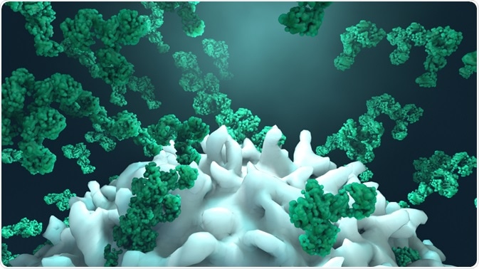
Antibodies are proteins manufactured by the B cells within the immune systems of higher life forms as an adaptive feature to allow the body to address invading foreign molecules that pose a threat to the organism. The natural function of antibodies involves their attachment to antigens (belonging to the invading foreign bodies), to flag them for destruction by the immune system.
 Image Credits: Design_Cells / Shutterstock.com
This innate propensity to attach themselves to specific molecules has established their use in scientific research. Because specific antibodies can be chosen that are known to latch onto certain antigens, researchers can use antibodies to identify the presence of certain substances. Consequently, the use of antibodies has become heavily relied upon in the study of protein function in cells.
Numerous techniques have been developed that exploit the nature of antibodies in order to investigate proteins in cells and tissues. Applications such as immunohistochemistry, immunocytochemistry, Western blot, ELISA, flow cytometry and FACS, and ELISPOT, are all established methods that utilize antibodies.
In order to make the most of these powerful investigative techniques, it is essential that the correct antibody is chosen that suits the specific application. Below we discuss how to choose the correct antibody for your application.
Choosing the right application
The first step of choosing the right antibody for your experiment is to ensure you select the correct application that is most suitable for your research purposes. Below, we outline the main features of the various methods:
Applications for studying localization
Immunohistochemistry has a low to medium throughput, can be used for fixed cells and tissues, can produce cellular localization, has a high spatial resolution, typically focuses on a single protein, can image multiple proteins at once, and is qualitative and semi-quantitative.
The other technique that studies localization is immunocytochemistry. This technique has a low to high throughput, can be used for fixed cells and tissues, can produce subcellular localization, it can image multiple proteins at once, and is qualitative and semi-quantitative. However, its spatial resolution is limited by tissue structure.
Applications for studying concentration
Western blot is the appropriate technique for studying protein concentration. It has a low to medium throughput, can be used for cell and tissue lysates, generates relative protein levels, can establish molecular weight, isoforms and post-translational modifications, and is semi-quantitative.
Applications for studying interaction
ELISA is the appropriate technique for studying protein interactions. The method has a medium to high throughput (96 well plate), it can be used to study serum/plasma, live or fixed cells and tissue, it produces accurate protein levels, it is quantitative and qualitative.
Applications for studying expression
ELISA can also be used for studying expression, its features are outlined above. Flow cytometry and FACS is the alternative technique of studying expression. It has a low to high throughput (up to 384 well plates). It can be used for fixed or live cells and can determine chemical and physical properties such as size, expression of surface markers, and more. It can be used to quantify and sort cells (FACS). Finally, it is a quantitative method.
Applications for studying identification
ELISPOT can be used when studying identification. It has a medium to high throughput (96 well plate) and can be used on live cells. It can identify specific cell(s) secreting an analyte of interest and has a high sensitivity. It can be both quantitative and qualitative.
Selecting the primary antibody
When selecting the primary antibody, the correct type, either monoclonal or polyclonal, must be considered.
Monoclonal antibodies recognize only a single epitope on the antigen, they generate less background noise as they are less likely to cross-react with other proteins. Also, their homogeneity is very high, meaning that the variability between batches is low.
They are also extremely efficient at binding with the antigen within a mixture of related molecules. However, they don’t work in a full range of species due to them being too specific. They also are vulnerable to epitope loss through chemical treatment of the antigen.
On the other hand, the features of polyclonal antibodies are as follows: They recognize multiple epitopes on any one antigen, they have a high affinity and can help amplify the signal from low expression proteins, they are more tolerant to minor changes to the antigen, and they can be used to detect proteins from species other than that of the immunogenicity. However, the downsides are that they are prone to batch-to-batch variability and can produce a background signal.
Selecting the secondary antibody
The type of method being used will inform which secondary antibody is suitable. Below we outline those types that are useable for technique.
- IHC: HRP or AP-conjugated, fragment, biotinylated (avidin/streptavidin conjugated to enzyme or fluorochrome), fluorochromes such as Alexa Fluor®, Cy® dyes, FITC, and PE, and pre-adsorbed.
- ICC: Fragment antibodies, biotinylated (avidin/streptavidin conjugated to fluorochrome), fluorochromes such as Alexa Fluor®, Cy® dyes, FITC, and PE, and pre-adsorbed.
- Western blot: HRP or AP-conjugated, biotinylated (avidin/streptavidin conjugated to enzyme), fluorochromes such as Alexa Flour® (Alexa 680 and Alexa 790), light chain specific antibodies VeriBlot for IP.
- ELISA and ELISPOT: HRP or AP-conjugated, biotinylated (avidin/streptavidin conjugated to enzyme), monoclonal (especially subtype-specific antibodies such as anti-IgG1)
- Flow cytometry and FACS: Fluorochromes such as Alexa Fluor®, Cy® dyes, FITC, and PE, biotinylated (avidin/streptavidin conjugated to fluorochrome), monoclonal, F(ab’)2 fragment, and conjugated primary antibodies for maximizing time efficiency.
Sources:
Dowd, S., Halonen, M. and Maier, R. (2009). Immunological Methods. Environmental Microbiology, pp.225-241. https://www.sciencedirect.com/science/article/pii/B9780123705198000122
Murata, L., Brunhoeber, P., Clements, J., ElGabry, E., Feng, J., Kapadia, M., Mistry, A., Singh, S. and Walk, E. (2019). Immunohistochemistry. Companion and Complementary Diagnostics, pp.53-91. https://www.sciencedirect.com/science/article/pii/B9780128135396000043
Pillot, J. (1996). The year of Pasteur: from the concept of antibody and antigen by Bordet (1895) to the ELISA. What future for immunological diagnosis?. Clinical and Diagnostic Virology, 5(2-3), pp.191-196. https://www.sciencedirect.com/science/article/pii/0928019796002218
Further Reading