Electron microscopy is a technique that uses a beam of accelerated electrons to illuminate and produce images of specimens. Using electron microscopy, much greater magnification levels and resolution can be achieved than with a light microscope because the wavelength of electrons is so much shorter than that of photons.
This enables scientists to study small specimens in fine detail and look at the ultrastructure of cells, microorganisms, crystals and metals, for example. Electron microscopes are much larger and more expensive instruments than the light microscope.
There are two main types of electron micrscope, namely, the transmission electron microscope and the scanning electron microscope.

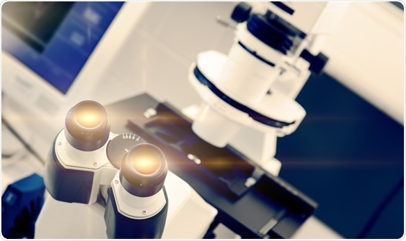
Laboratory Electron Microscope - Image Copyright: science photo / Shutterstock
The Inventors of the Electron Microscope
It was in 1931 that the German scientists Max Knoll and Ernst Ruska developed the first model of an electron microscope. Ruska followed this with another prototype two years later that could provide a greater resolution than the light microscope, an achievement for which he received a Nobel prize fifty years later.
The Differences Between a Transmission Electron Microscope and a Scanning Electron Microscope
A transmission electron microscope produces an image by passing a high voltage electron beam through a very thin specimen that is semi-transparent to electrons. The beam that is transmitted through the specimen carries structural information about the specimen, which can then be magnified by the microscope. By contrast, a scanning electron microscope detects secondary electrons that arise from the surface as a result of excitation by the original electron beam.
The scanning electron dicroscope does produce lower resolution images than the transmission electron microscope, but since it uses surface electron interactions, it can image bulk samples and provide a much greater depth of field. This enables scientists to achieve 3D information about the physical features of the specimen.
Scanning transmission electron microscopy combines the technology of both the scanning electron microscope and the transmission electron microscope and can be performed using either piece of equipment. Similarly to transmission electron microscopy, samples need to be very thin as the technique mainly involves looking at the electron beam transmitted through and emerging from the specimen.
Components
Electron microscopes include the following components:
- An electron gun to produce and accelerate the electrons
- A series of magnetic lenses to help direct and focus the electron beam
- An objective lens, which produces the first, intermediate image
- A vacuum system to ensure electrons do not collide with molecules of air
- An image capturing system, which may be a charge-coupled device (CCD) camera, an imaging plate or negative film.
Applications of Electron Microscopy
Electron microscopy has many wide ranging applications in science and technology.
It can provide high resolution information about cells, organelles, tissues, microorganisms, biopsy samples, macromolecules, crystals and metals.
It has been used to study pathogens such as viruses and bacteria and is particularly valuable in the study of emerging diseases and germs used in bioterrorism. It has previously been used as a technique in the diagnosis of diseases such as smallpox, hepatitis B, gastroenteritis and parvovirus B19.
Electron microscopy also has applications in the electronics industry where it is used to control manufacturing processes. The technology is used worldwide in various other industrial applications including aeronautics, the manufacture of motor vehicles and the clothing industry, to name but a few.
In forensics, the technique is used for the analysis of clothing fibres and blood and gunshot residues, for example.
References
Of Interest
Clinical Microbiology Reviews Journal, Modern uses for electron microscopy in virus detection:: http://www.ncbi.nlm.nih.gov/pmc/articles/PMC2772359/
Further Reading