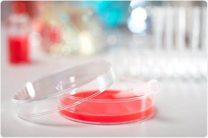
In order to understand how cells associate to form tissues and organs, it is necessary to determine the mechanisms that drive cell behaviors. Rather than studying cells in vivo, which would be significantly difficult, scientists use cell cultures. These are cells that have been removed form an organism and cultured in the laboratory.
 anyaivanova | Shutterstock
Using cell cultures, scientists are able to manipulate the cellular microenvironment in order to simulate physiological conditions in vivo.
Which type of cell culture is best for manipulating cells in vitro?
For over a century, two-dimensional (2D) cell cultures were used to study cellular response to biophysical and biochemical cues. While 2D cultures have greatly advanced our understanding of cell behavior, they have their own limitations. For example, cells in culture demonstrate bioactivities that differ from those observed in vivo. Thus, research results may not be fully representative of in vivo reactions.
To address this problem, novel 3D cell culture scaffolds are being developed, particularly as they accurately mimic conditions in vivo. These are referred to as spheroid or organoid cultures and have demonstrated success in inducing in vivo cell behaviors fates when stimulated by particular processes or cues under study.
However, 3D models come with several challenges. These include creating an interface between the different tissue types; controlling the distribution of bioactive factors (e.g. hormones and chemical messengers) spatially and temporally, and maintaining the passage nutrients and gases necessary for cell survival.
When faced between the choice of 2D or 3D systems, the specific process of interest under study must be considered. Whilst 3D platforms are better at inducing in vivo -like cell behavior, they are limited by a lack of universal application; at present, only some cell types can be grown in this way. 2D cell cultures thus offer a workable, easy to use alternative.
Current 2D cell culture methods
Culturing cells requires a flat surface to which the cells can adhere and grow. Typical materials used for this purpose include glass or polystyrene. Owing to the uniform nature of the platform, cells grow homogenously as they have access to similar concentrations of nutrients and growth factors present in the medium. This uniformity makes 2D cultures an attractive option in clinical and research settings.
Despite this simplicity, 2D cultures do not allow control of cell shape – which is a critical determiner of response to biophysical cues in vivo. To account for this, micro-patterned substrates such as cell-adhesive islands, microwells, and micropillars have been designed to aid in cell shape customization.
These solutions offer a pseudo-3D environment that can stimulate apical-basal polarity, which refers to the asymmetry in the organization and distribution of cellular components. This can be problematic when apical-basal polarity may be unnatural in vivo.
Induced polarity may, therefore, be favorable as it enables the native cells to perform their natural functions (migrating, distributing and sensing environmental cues). The effect of unwanted polarity can be mitigated by using a sandwich culture methodology.
In this technique, an additional platform is placed on top of the uniform layer of cells, providing them with the same extracellular matrix (ECM) proteins that coat the supporting bottom layer. This effectively prevents cell polarization, providing a mimic of the 3D environment seen in vivo.
Current 3D cell culture models
The third dimension made available in 3D cultures enables the accurate modeling of in vivo environments in vitro. This provides greater structural complexity to cells, allowing them to maintain a steady (homeostatic) state across a greater period of time. Subsequently, cell cultures grown using 3D culturing methods are more predictive of what may happen to cells in vivo than those grown in 2D.
Some 3D culturing methods include microfluidics. This refers to the manipulation of small fluid volumes within artificially fabricated microsystems. Soluble factors that regulate biochemical behaviors are well dispersed in microfluidic cell cultures, which closely mimic their delivery in vivo.
Another benefit of microfluidic 3D cell cultures is the ability to link different tissue types together and examine how they interact. Consequently, researchers are able to better understand how organs and systems, and not just cells and tissues function.
Furthermore, the flow of fluids (such as interstitial fluids and blood) is important for cell function, especially as cell differentiation and metabolism is dependent on this. Therefore the control of fluid pressures enabled by microfluidic techniques is invaluable.
Another benefit of 3D cultures is improved modelling of barrier tissues. In vivo, epithelia form a barrier between organ compartments. The barrier function is important as it regulates the affect that environmental variables have on the underlying compartment. The proper function of the epithelial barrier is crucial for survival, and so its representation in 3D cultures improves the success of integrated organ systems in vitro.
Comparison of cell development in 2D and 3D environments
The prevalence of 3D culture techniques is increasing as advances allow more accurate mimicry of in vivo conditions. 2D cultures are limited by their inability to appropriately influence cell-cell interaction, cellular mechanics, and access to nutrients.
Despite this, 2D cultures are successful in epithelial systems; for example, lung airway epithelia will develop normally in vitro. The main drawback of 2D systems are their simplicity, often failing to cause cultured cells to demonstrate the expected cell development processes. Ultimately, their simplicity presents a disparity between the biologically relevant information that is required and what is provided in 2D.
3D cultures are similarly limited. The throughput, which is the total output the 3D system can produce, is low compared to 2D methods. Many techniques are time-consuming and difficult to set up.
Consequently, they cannot be used in drug development which relies on large numbers of screening measurements to be taken. 3D cultures are also limited by their incompatibility with forms of microscopic analysis. This problem stems from the large size of 3D cell cultures; this also prevents proper distribution of nutrients to cells, both at the correct location and time.
Considering that 3D cell culture methods are relatively new and emerging, the potential of 3D cell cultures is promising. Their ability to accurately recapitulate the in vivo cell environment makes them the preferred method of choice in the clinical and research setting.
Further Reading