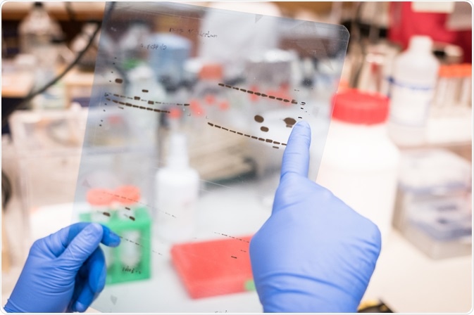
Western blot assays are a popular technique used to analyze protein expression by determining the molecular weight of proteins, their post-translational modifications, and their abundance.
 SINITAR | Shutterstock
The key to a successful Western blot is achieving a signal to noise ratio allowing the detection of proteins while minimizing the background noise. High background noise can have several causes and associated solutions.
Detection Issues
Selecting the right antibody
A uniform high background is when the entire blot is darkened, which results in it being harder to see specific bands. Detection issues can be due to high concentrations of antibodies, which leads to saturation and coating of the membrane and thus nonspecific binding. This can be solved by reducing the antibody concentration via a dot-blot test to find the optimal antibody concentration.
Exposure time
Another problem related to detection issues that can lead to high uniform background noise is when the membrane is overexposed. If the exposure times are increased, it can enhance the overall background without increasing the sensitivity. This can be fixed by adjusting the exposure parameters and using a shorter exposure time.
Selecting a good quality membrane
Other solutions include loading more proteins, with the aim of increasing the target antigen’s abundance, titrating the primary antibody well and trying a different primary antibody if there are several alternatives available.
Other issues may be caused by the membrane itself. Certain membranes are considered to give more background noise than others. For example, nitrocellulose membranes are generally thought to give less background than polyvinylidene difluoride membranes(PVDF). However, PVDF membranes have a high binding capacity, while nitrocellulose membranes tend to be brittle and often do not bind smaller proteins.
Membrane selection should be done based on empirical data for each antigen-antibody combination. Furthermore, a dried out or old membrane that has passed its expiration date can contribute to high background.
Antibody storage
Sometimes, the binding activity of the antibody can be insufficient. This generally occurs if the handling and storage of antibodies was inappropriate. For example, thawing and repeatedly freezing antibodies should be avoided.
Each time a Western blot is prepared, a fresh aliquot of antibody that has been stored at -20 °C should be used. If antibodies are to be stored for long periods, it is recommended that they be stored at -80 °C to remain stable.
Blocking Issues
Uniform high background can also be caused by blocking issues, specifically insufficient blocking of the membrane.
Membrane blocking stops nonspecific binding of the antibody to the membrane and is done by incubating the blot with a blocking buffer. As such, both the temperature and length of the incubation can affect the success of membrane blocking and both can be increased in the event of insufficient membrane blocking.
Standard blocking is done for 1 hour at room temperature with agitation, or overnight at 4°C with agitation. Furthermore, the type and concentration of blocking reagent can affect its success. The type of blocking reagent to use depends on the interaction between the antigen and antibody, but a common initial choice is non-fat dry milk.
Typically, it is recommended to start at 1% or 5% concentrations of milk. If the antibody or detection system is incompatible with milk, 3% bovine serum albumin (BSA) is often used.
Another blocking issue that can lead to uniform high background are reactions between blocking agents and antibodies, resulting in interference. If the blocking agent is protein based, antibodies can bind to those proteins and cause high uniform background. For example, animal serum with unpurified primary antibodies can bind to animal proteins in blocking reagents.
As mentioned previously, certain blocking reagents are unsuitable for certain detection systems – milk contains biotin, and can react with avadin when an avadin-biotin detection system is used. If neither non-fat dry milk nor bovine serum albumin are suitable, protein-free or non-animal protein blocking reagents are available from some retailers. Alternatively, a secondary control lacking primary antibody can be run.
Contamination issues
Lastly, contamination can cause high background in Western blot assays. Contamination can occur when blocking solutions are reused, which they should never be. An alternative is to use pure protein as a blocking agent. Furthermore, buffers which contain detergent and milk can promote bacterial growth and should, therefore, be made fresh to avoid bacterial contamination.
Further Reading