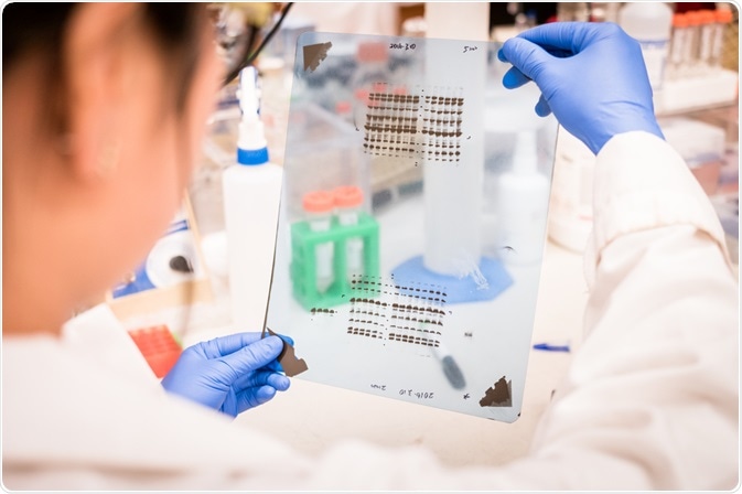
Immunohistochemistry and Western blotting both work by exploiting the principle of how antibodies specifically bind to the antigens present in biological tissue.

Image Credit: Sinitar/Shutterstock.com
Immunohistochemistry is the most common of immunostaining techniques. It visualizes the presence of targeted antigens within a tissue sample by using antibodies that attach themselves to the antigens, having a catalytic effect, and splitting the molecule into identifiable compounds. Visualizing this interaction can be achieved by using an enzyme (producing a chromogenic signal) or a fluorophore (creating a fluorescent signal).
Western blot works on the same principle. It was first reported back in the 1970s when scientists first used it to successfully classify different proteins existenin biological tissue. Since then, significant advancements in the technology have been made, leading to the establishment of the modern fluorescent, colorimetric, and chemiluminescent Western blot technique.
All versions of the Western blot method rely on the workings of the antigen-antibody complex to identify specific proteins present in a sample, indicated by either the enzyme-substrate reactions of colorimetric, chemiluminescent, or fluorescent molecules. The light produced by these sources can be used to infer the presence and concentration of the proteins in a sample.
Comparing the techniques
While the two methods rely on the principle of the antibody-antigen interaction, there are some major differences between the two techniques. Below, we break this down, step by step.
Firstly, for both techniques, the sample must be correctly prepared. The preparation differs quite significantly between the two. For immunohistochemistry, the tissue samples are sliced or left whole, depending on their size.
The limit for the size of samples generally ranges from 3 µm to 5 µm. Next, samples are embedded into a medium, usually paraffin wax or cryomedia. Following this, the slices are placed on slides and dehydrated using increasing strengths of alcohol washes, finally, ending with a detergent being added to clear the sample, making it ready to be viewed under a microscope.
On the other hand, Western blot requires that samples are first separated by electrophoresis and then immobilized in a blotting membrane.
The method of sample staining is also slightly different between the two techniques. Immunohistochemistry adds either polyclonal or monoclonal antibodies. These antibodies are classed as either primary or secondary reagents, the first being those raised against a target antigen and are usually unconjugated, the latter is conjugated to a linker molecule that then recruits reporter molecules.
The conjugated version is used in a direct staining method, where the antibody directly reacts with the antigen in the sample, the unconjugated version is used in an indirect staining method where the unlabeled antibody binds to the target antigen within the tissue, causing a secondary antibody to react with the primary antibody.
In comparison, the Western blot method sees a fluorescent dye added to the sample, followed by exposure to a light source of the appropriate wavelength to excite the molecules of the fluorophores.
After some time, the excited fluorophores eventually release the energy that they gained from the light source to return to their ground state. This release of energy is what makes the fluorophores fluoresce, emitting a light source that can be captured by a digital imager.
Comparing the benefits and drawbacks
The main advantage of immunohistochemistry is that the method allows researchers to detect the exact location of a target protein within a tissue sample. This main advantage has led to it becoming well established within the field of neuroscience as a robust method for investigating protein expression in target brain locations.
On the other hand, the method has a major disadvantage when compared with the Western blot method. This is the fact that the stains are not checked against a molecular weight ladder, as they are in Western blot, which means that there is no way to prove that the staining shown in immunohistochemistry is conclusively related to a specific protein.
The indirect method of immunohistochemistry is recognized for being highly sensitive, however, the direct method is known for being the opposite, and is generally incapable of detecting small concentrations of target antigens.
The Western blot method is advantageous in that is has established itself as a reliable technique for gathering quantitative data. However, its most prominent advantage is that it has proven to be effective at generating a signal that is proportional to the amount of the protein that exists in the sample.
Also, the Western blot is often chosen for its ability to detect numerous target proteins simultaneously, having the effect of significantly cutting down on testing time as well as reducing the number of resources required.
A difference in applications
There is a great deal of overlap in the applications of the two techniques given that they are so similar. However, overall, immunohistochemistry is mostly relied on as a diagnostic tool for numerous cancers.
The method is used to identify abnormal cells that are characteristic of certain tumors. The method is also widely used to identify and locate proteins and biomarkers found in both normal and diseased tissues. Finally, it is also commonly used to detect numerous infectious organisms within tissues.
In comparison, the Western blot is mostly used for the detection of autoimmune diseases, allergies, and infectious diseases. It is widely used in the fields of molecular biology, biochemistry, and cell biology, with its most notable applications being used as a diagnostic tool for HIV and BSE.
Sources:
Ditaddi, R., Catozzi, L., Gion, M., Brazzale, A., Capitanio, G., Gelli, M., Menegon, A., Gardini, G., Malagutti, R. and Piffanelli, A. (1993). Comparison between western blotting, immunohistochemical and ELISA assay for p185neu quantitation in breast cancer specimens. Anticancer Res, 13, pp.1821-4. https://www.ncbi.nlm.nih.gov/pubmed/7903522
Kaliyappan, K., Palanisamy, M., Duraiyan, J. and Govindarajan, R. (2012). Applications of immunohistochemistry. Journal of Pharmacy and Bioallied Sciences, 4(6), p.307. https://www.ncbi.nlm.nih.gov/pmc/articles/PMC3467869/
Lund-Johansen, F. and Browning, M. (2017). Should we ignore western blots when selecting antibodies for other applications?. Nature Methods, 14(3), pp.215-215. https://www.nature.com/articles/nmeth.4192
Najafov, A. and Hoxhaj, G. (2017). Introduction. Western Blotting Guru, pp.1-3. https://www.sciencedirect.com/science/article/pii/B9780128135372000011
Towbin, H. (1998). Western Blotting. Encyclopedia of Immunology, pp.2503-2507. https://www.sciencedirect.com/science/article/pii/B0122267656006496
Further Reading